 NA-MIC Project Weeks
Back to Projects List
Establishing Feature Correspondences between ultrasound images acquired at different time points during brain tumor resection
Key Investigators
- Nick Jowkar (Brigham and Women’s Hospital)
- Clement Mirabel (Brigham and Women’s Hospital)
- Sarah Frisken (Brigham and Women’s Hospital)
- Tina Kapur (Brigham and Women’s Hospital)
Project Description
The overarching goal of this project is to use pre- and intra-operative imaging to help neurosurgeons determine whether gross total resection has been achieved.
Objective
- Objective A. Manually identify 10-15 corresponding features in ultrasound images from 5 patients. Save results.
- Objective B. Review these landmarks with available neurosurgical ultrasound experts at project week
Approach and Plan
- We will identify corresponding landmarks or features in 3D ultrasound images acquired during different time points in brain tumor resection.
- We are using 3D Slicer to visualize, explore, and annotate these landmarks in ultrasound images.
Progress and Next Steps
- Annotated intra-operative ultrasound for 4 patients.
- Next step - measure intra-operator and inter-operator variability in these landmarks
Illustrations
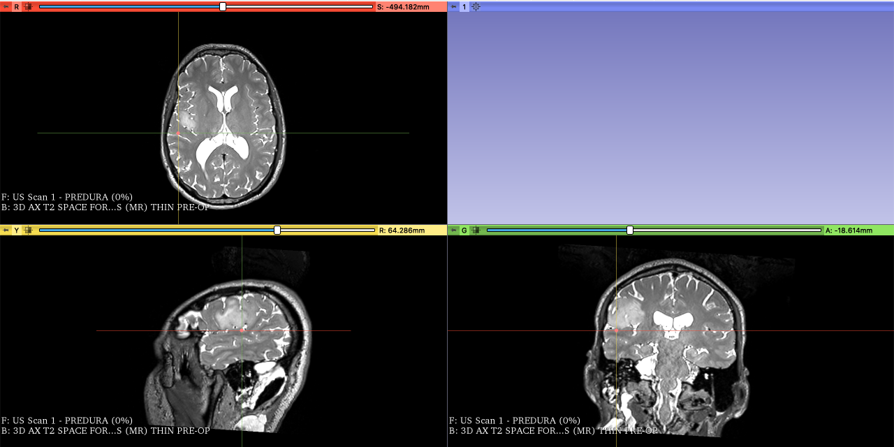
- Picture of the fiducial on the T2 pre-operative MRI image located on a sulcus
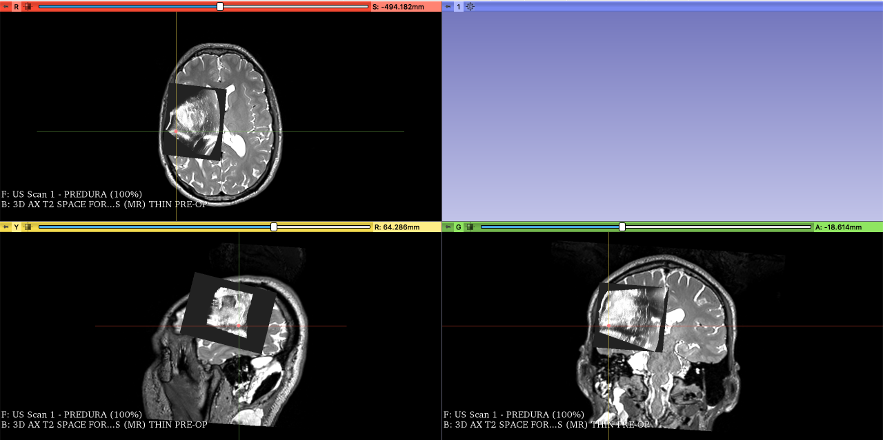
- Picture of the pre-dura Ultrasound scan on top of the MRI scan showing the same fiducial on the sulcus
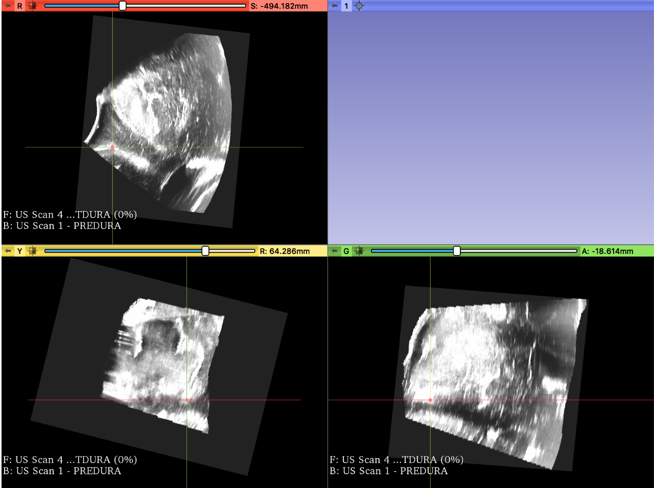
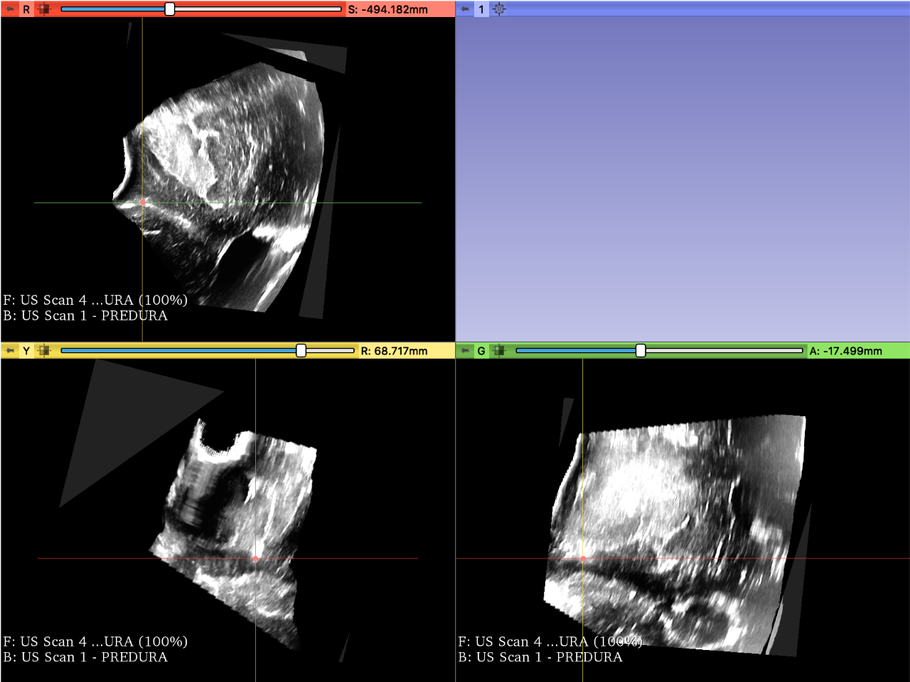
- Screenshot of the pre-dura ultrasound with the fiducial on the sulcus
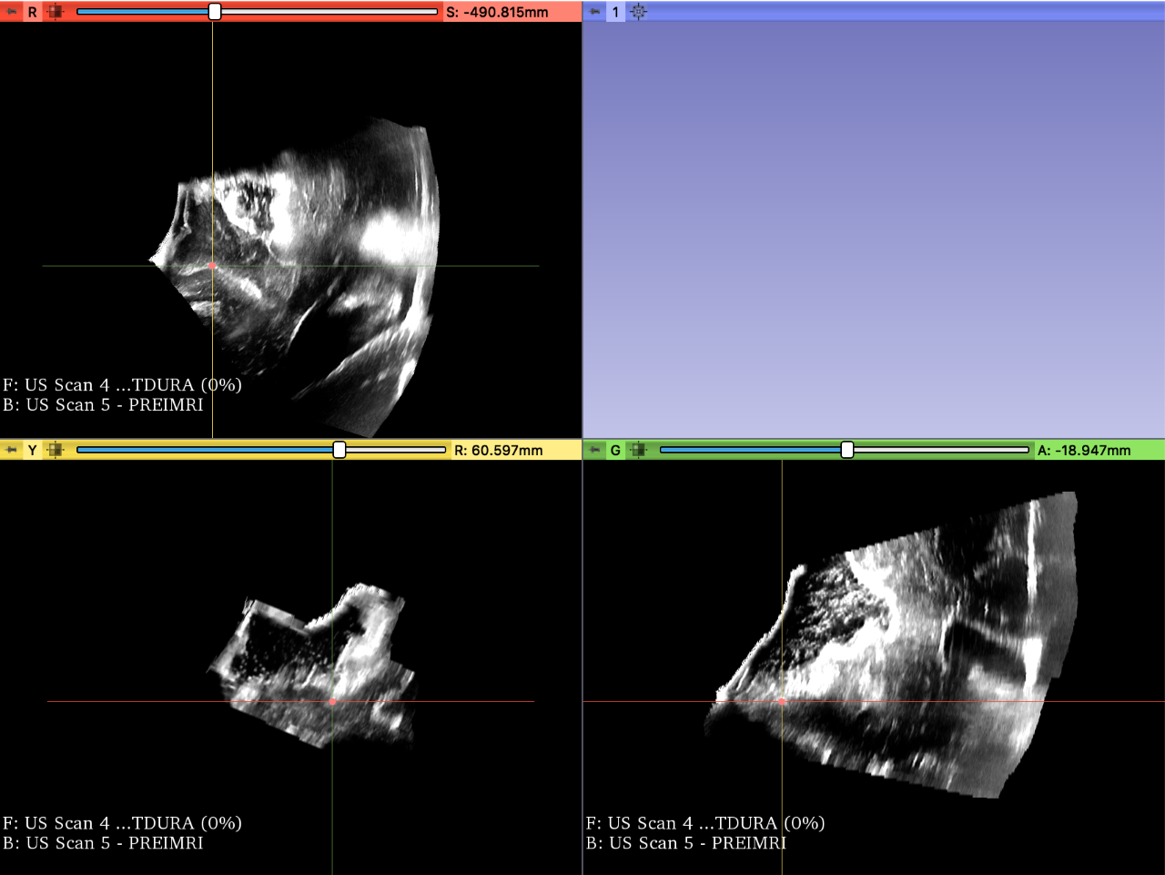
- Screenshot of the post-dura ultrasound fiducial located on the sulcus
- Screenshot of the pre-iMRI of the fiducial located on the sulcus